
Synthetic biology (or SynBio) is an exciting intersection of biology and engineering, which promises to t…
Change your region and language
Be sure to pick the location that matches your preferences.
Benefits can be reinvested in accelerating current or upcoming projects, used to hire new staff or support your overall growth. Finding out if you qualify is quick and easy.
To improve the competitiveness of the UK’s most energy intensive businesses, the Government implemented a number of compensation schemes to mitigate the impact of renewables policies, including Energy-Intensive Industries (EII), UK ETS Compensation (UKETS) and Climate Change Levy (CCL).
A better way to obtain valuable savings on capital spend. We can help your company obtain valuable cash tax savings and improved business cash flow. The UK government allows businesses who pay tax in the UK to claim capital allowances on qualifying capital expenditure they incur, when they buy equipment or buildings, carry out new construction, refurbishment works or fit out works.


Our tax and technical experts help thousands of businesses each year to simplify their access to complex incentives and maximize their financial benefit. In the past year alone, we have helped our UK clients successfully claim more than £900m in R&D Tax Credits to support their growth plans.
Our in-house team of highly qualified tax and technical experts are committed to help you leverage innovative strategies tailored to your unique business environment. We have 25+ years of experience working with businesses of all sizes offering tax services.
We help businesses to claim significant tax relief from innovative projects.
We identify eligible businesses that can apply for compensation for a significant proportion of the costs of their energy bills.
Our Land Remediation team leverages their proven experience in this area to offer a bespoke consultancy service.
We can help your business obtain valuable cash tax savings and improved business cash flow.
Patent Box can reduce your corporation tax rate on qualifying IP income to 10%
We help businesses to benefit from four valuable creative tax reliefs – Video Games, Museum and Galleries Exhibition, Theatre and Orchestra.
With more than 12 years of R&D experience and unrivalled tax and technical expertise. Leyton UK is ideally positioned to unlock potential value within your business
Read more Case Studies arrow_forward
Alvis Brother are continuously striving to minimise the environmental impact of its farming practices via conducting responsible farming techniques. This is a key part of the company’s ethos, and as such, the company’s R&D team underwent significant development work in attempts in improve the sustainability of their practices. Find out more about the work Alvis Brothers are undertaking.


Find out how Seanamic Group were able to increase their R&D Tax Credit claim value by 400% when claiming with Leyton.

Find out how FGP Systems were able to optimise their previous R&D Tax Credits claim with Leyton.

Find out how Leyton helped Alex Thomson Racing claim R&D Tax Credits on their cutting edge projects.
Take some time with one of our experts to see if you are entitled to tax relief.
Get an estimate of how much we can secure for you, by filling out the simple form on our contact page.
Find out today 🡪
Our expert team will resolve your HMRC R&D enquiry, protecting your business and securing the best outcome for you.
Check our Enquiry Defense Support service 🡪
Synthetic biology (or SynBio) is an exciting intersection of biology and engineering, which promises to t…

Even in the best of circumstances, we know that it can be challenging to balance a successful career and …

Small and Medium-sized enterprises (SMEs)are the lifeblood of innovation in the UK. We’ve dived into the …

Full expensing is a first-year allowance that allows businesses to reduce their tax liability and free up…

To help businesses understand if their work qualifies for R&D Tax Relief and to make sure that compan…

Software development is essential for driving some of the UK’s most exciting industries, such as cybersec…
At Leyton, we believe in empowering our employees to thrive and grow. Our career development programs present dynamic opportunities to gain positive mobility in your career. Gender equality, practice that promotes diversity, and engaging company culture are key elements of Leyton’s values as an employer.
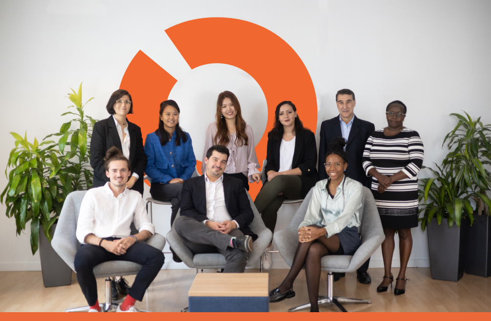
As a business, we want to help unleash our clients’ potential and be a strategic partner in their evolution and growth. We help find openings in processes and ways of working that others can’t see, then unlock these opportunities to create value efficiency.
Contact our Experts arrow_forward arrow_forward